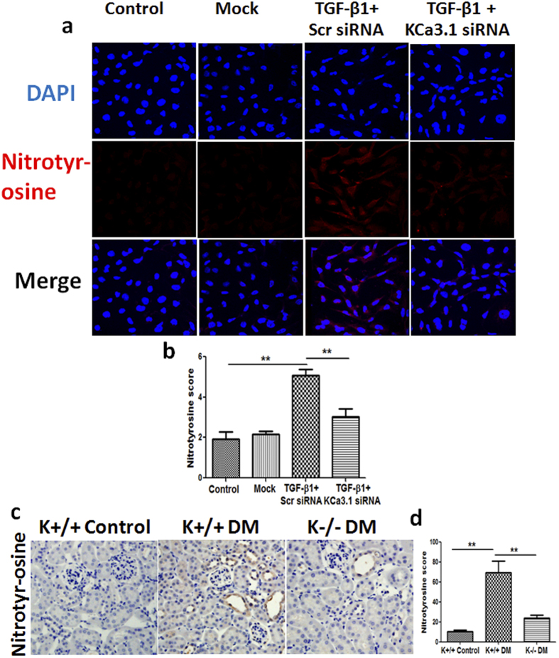
Figure 8. Effect of knockdown KCa3.1 on the expression of nitrotyrosine in TGF-β1-stimulated HK2 cells and the expression of nitrotyrosine in STZ-induced diabetic KCa3.1+/+ and KCa3.1−/−mice.
HK2 cells transfected with scrambled siRNA or KCa3.1 siRNA were untreated or treated with TGF-β1 for 48 h. Immunofluorescence staining of nitrotyrosine in TGF-β1-exposed HK2 cells transfected with or without KCa3.1 siRNA (a). Quantification of nitrotyrosine expression in TGF-β1-exposed HK2 cells (b). N = 4. KCa3.1+/+ and KCa3.1−/− mice were injected with STZ to induce diabetes or citrate buffer alone as non-diabetic control. After 24 weeks diabetes, kidney tissues were collected for immunostaining. Immunohistochemical staining of nitrotyrosine in mice kidney tissues (c). Quantification of nitrotyrosine expression in mice kidney tissues (d). N = 8. Results are presented as mean + SEM. *P < 0.05 and **P < 0.01. Original magnification: ×600.

