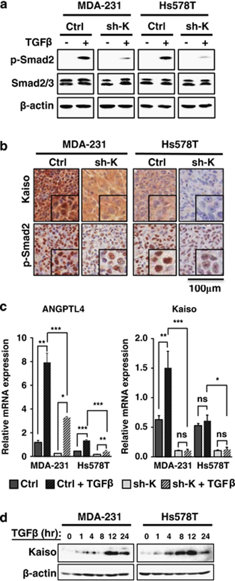
Figure 4.
Kaiso-depletion attenuates TGFβ signaling and transcriptional responses. Cells were treated with 10 ng/ml of TGFβ for 1 h before assaying for TGFβ activity. (a) TGFβ treatment of control MDA-231 and Hs578T cells results in increased p-Smad2 levels. However, Kaiso-depleted MDA-231 and Hs578T cells treated with TGFβ display reduced p-Smad2 levels. (b) Kaiso-depleted MDA-231 and Hs578T xenografts exhibit decreased TGFβ signaling as evidenced by reduced p-Smad2 protein levels. (c) TGFβ-induced expression of ANGPTL4 is attenuated in Kaiso-depleted cells treated with 10 ng/ml of TGFβ for 24 h. Interestingly, Kaiso expression is significantly increased by TGFβ treatment in MDA-231 cells. (d) Immunoblot analysis revealed a peak in Kaiso protein levels at 12 h in both MDA-231 and Hs578T cells in response to TGFβ treatment. All experiments were performed in triplicate. Representative images from all experiments are shown. *P<0.05, **P<0.005, ***P<0.0001, NS, not significant. β-Actin serves as a loading control.

