
Fig. 1.
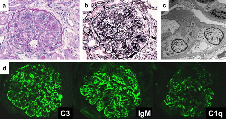
First biopsy of patient 2: a (PAS stain, magnification ×400); b (PAM stain, magnification ×400), mesangial proliferation, increased lobulation, doubled contours: c (electron microscopy), subepithelial, subendothelial, mesangial deposit: d (immuno-fluorescence microscopy), C3 (++), IgM (++), C1q (+), IgG (+) fringe pattern