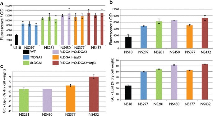
Fig. 4.

Comparison of lipid accumulation in Y. lipolytica strains with different target combinations by different methods. a Strains analyzed by fluorescence assay after 96 h of fermentation in a 48-well plate. Two or three transformants were analyzed for each construct and average with standard deviation is shown. b Strains analyzed by fluorescence assay and GC after 96 h of fermentation in 50-mL flasks. The measurement was done in triplicates and average with standard deviation is shown. c Strains analyzed by GC after 140 h of fermentation in 1 L bioreactors. With exception for NS450 the measurement was done in duplicates and average with standard deviation is shown
