Figure 1.
Phenotypic Diversity in Wild-Type Strains.
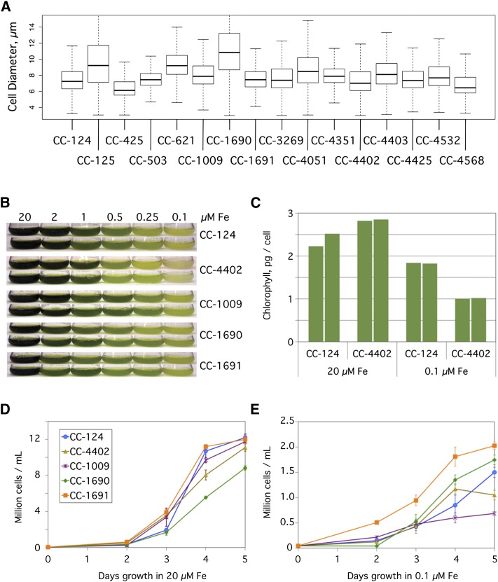
(A) Diversity of cell size. The size of Chlamydomonas cells from the indicated 16 strains were assayed by a cellometer. Results are plotted as a box plot indicating the median cell size (bold horizontal line), the upper and lower quartiles (ends of boxes), and the range (thin horizontal lines) from 1000 cells per sample.
(B) Growth of Chlamydomonas cells in a range of iron concentrations. The indicated strains were inoculated to a density of104 cells/mL in 100-mL cultures of TAP media containing iron concentrations ranging from 0.1 to 20 µM. Duplicate cultures were photographed after 5 d of growth.
(C) Chlorophyll content in Chlamydomonas cells. Chlorophyll content was measured on a per cell basis for duplicate cultures of the closely related CC-124 and CC-4402 strains in TAP medium plus 20 µM or 0.1 µM iron after 5 d of growth.
(D) and (E) Growth rates. Quantification of the number of cells in TAP medium supplemented with 20 µM iron (D) or 0.1 µM iron (E). Cells were counted by a hemocytometer daily for 5 d and plotted. Each point represents the mean (± range) of the cell count for duplicate cultures.

