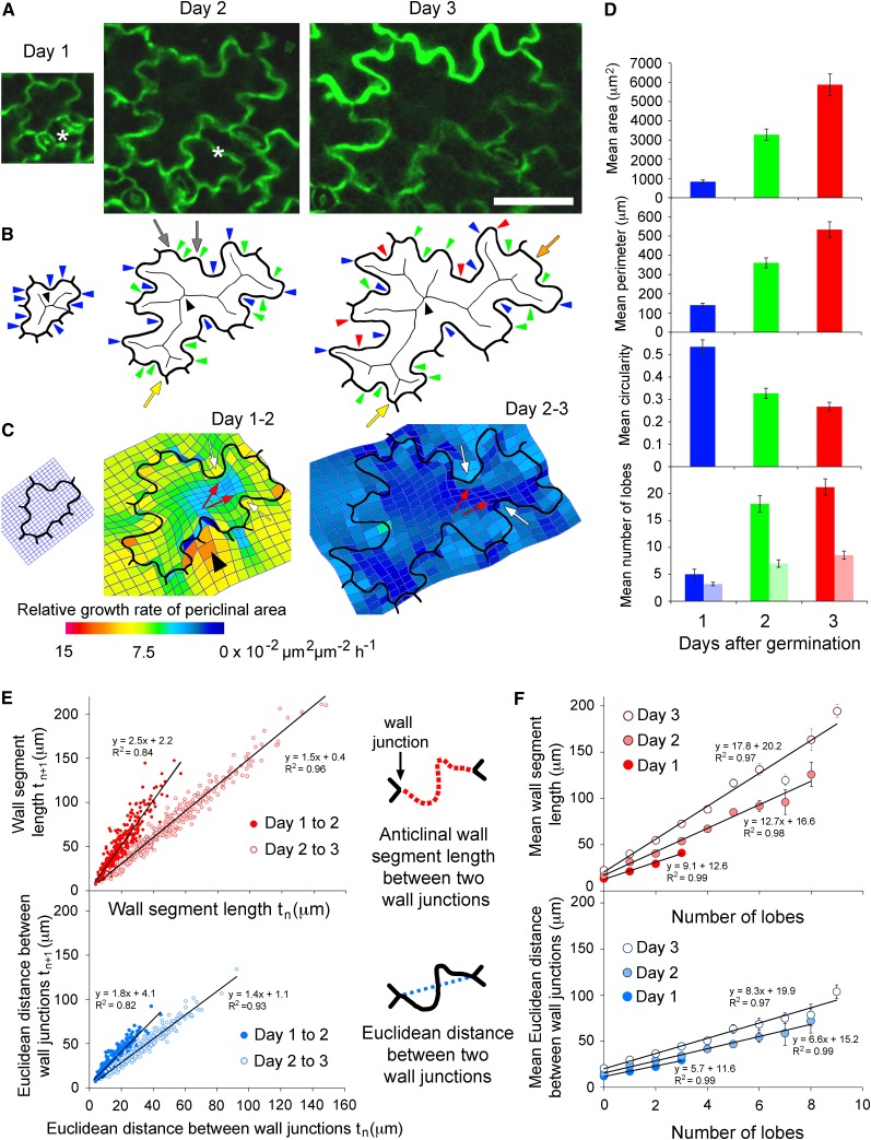
Figure 1.
Lobe Development and Growth of Arabidopsis Pavement Cells.
(A) A representative cell at 1, 2, and 3 d after germination. A neighboring cell divided between days 1 and 2 (asterisks). Bar = 50 µm and applies to all three images of the cell between days 1 and 3.
(B) Digital outlines of the cell. Lobes at day 1 (blue arrowheads) and new lobes at day 2 (green arrowheads) and day 3 (red arrowheads) were identified manually. Alternatively, concave lobes were identified at the branch ends of each skeleton (black arrowheads indicate the skeletons). Skeleton branch ends were sometimes present at wall junctions (yellow arrows) or regions of straight wall (orange arrow) and were not always located at small concave lobes (gray arrows).
(C) Thin-plate spline analysis (mesh overlying cell outline) of differential growth in cell walls between days 1 and 2 and days 2 and 3. Periclinal wall growth was predicted to be isotropic on the concave side of lobes (white arrows) and anisotropic on the convex side (red arrows). The region of intense growth (arrowhead) corresponds to the newly divided cell (asterisk in [A]).
(D) The mean area, perimeter, and circularity of 50 pavement cells at days 1, 2, and 3. A circularity value of 1 is a perfect circle, and 0 is an infinitely complex shape. The mean number of lobes (concave and convex) within each cell per day was identified manually (dark bars) or the mean number of concave lobes was ascertained by counting skeleton branch ends (light bars). Error bars are se of the mean.
(E) The length of each anticlinal wall segment between two wall junctions within the 50 pavement cells was compared between successive days. For each wall segment, the Euclidean distance between the two wall junctions was also compared between days.
(F) The number of lobes per mean anticlinal wall segment length between wall junctions or per Euclidean distance between wall junctions on day 1, 2, or 3. Error bars are se of the mean. The sample sizes on day 3 are: 154 anticlinal wall segments with 0 lobes, 116 with 1 lobe, 46 with 2 lobes, and 10 with 3 lobes; on day 4: 16 anticlinal wall segments with 0 lobes, 72 with 1 lobe, 77 with 2 lobes, 72 with 3 lobes, 48 with 4 lobes, 22 with 5 lobes, 12 with 6 lobes, 4 with 7 lobes, and 3 with 8 lobes; on day 5: 12 anticlinal wall segments with 0 lobes, 45 with 1 lobe, 71 with 2 lobes, 80 with 3 lobes, 57 with 4 lobes, 28 with 5 lobes, 19 with 6 lobes, 4 with 7 lobes, 7 with 8 lobes, and 4 with 9 lobes.

