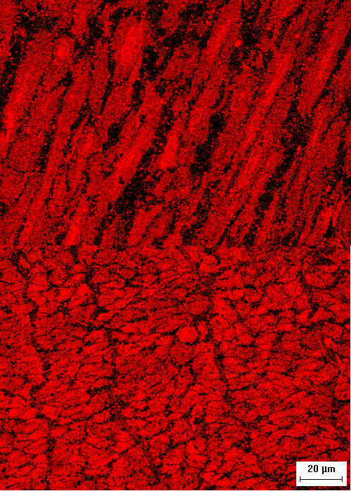
Figure 1.

Intracellular localization of HSP20 in swine carotid arteries. Representative cross-sectional (top) and longitudinal (bottom) confocal micrographs showing the distribution of HSP20 immunostaining in 10 μM histamine and 10 μM nitroglycerin treated swine carotid artery. Orientation refers to the long axis of the cells. The image is 160 microns wide. The micrographs show that HSP20 immunostaining was present throughout the cell, however, there were regions with more intense staining.
