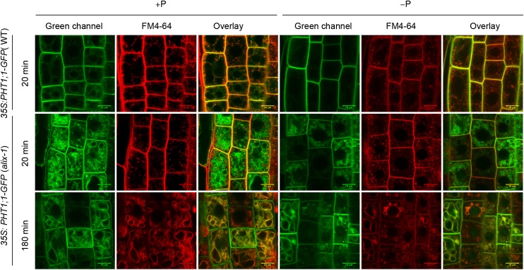
Figure 7.
PHT1;1-GFP Is Mislocalized in alix-1 Mutants.
Confocal images of root epidermal cells from 5-d-old wild-type (WT) and alix-1 seedlings overexpressing a PHT1;1-GFP fusion grown in +P and −P conditions. Seedlings were treated with 2 μM FM4-64 for 5 min, washed, and visualized after 20 and 180 min. The green and red channels correspond to the PHT1;1-GFP and membrane-associated FM4-64 fluorescence, respectively. Overlay of both channels in images after 180 min shows PHT1;1-GFP localization in alix-1 tonoplasts. Bars = 10 μm.

