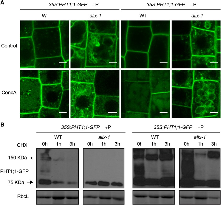
Figure 8.
alix-1 Mutation Alters PHT1;1-GFP Degradation.
(A) Confocal images of root cells from 5-d-old 35S:PHT1;1-GFP seedlings in wild-type (WT) and alix-1 mutant backgrounds treated with 1 μΜ ConcA for 6 h. Bars = 5 μm.
(B) Immunoblots showing PHT1;1-GFP (arrow) degradation over time in 10-d-old 35S:PHT1;1-GFP seedlings in wild-type and alix-1 mutant backgrounds grown in +P or −P conditions, incubated or not during 1 and 3 h with 50 μM cycloheximide. Anti-GFP was used to detect PHT1;1-GFP. Ponceau staining of the large subunit of Rubisco (RbcL) was used as loading control. An asterisk indicates the position of PHT1;1-GFP aggregates as previously reported by Bayle et al. (2011).

