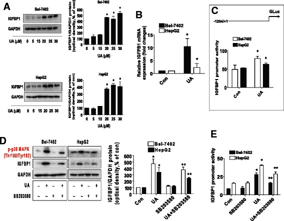
Fig. 3.

UA induced the protein, mRNA expression, and promoter activity of IGFBP1, which were blocked by SB203580. a-b, HepG2 and Bel-7402 cells were exposed to increased concentrations of UA or UA (25 μM) for 24 h. Afterwards, the expression of IGFBP1 protein (a) and mRNA (b) were detected by Western blot and qRT-PCR methods as described in the Materials and Methods section. *Indicates significant difference as compared to the untreated control group (P < 0.05) c, Bel-7402 and HepG2 cells were tranfected with wild type human IGFBP1 promoter reporter construct ligated to luciferase reporter gene and internal control secreted alkaline phosphatase (SEAP) for 24 h, followed by treating with UA (25 μM) for an additional 24 h. Afterwards, the IGFBP1 promoter activity were detected by the Secrete-Pair Dual Luminescence Assay Kit. d, HepG2 and Bel-7402 cells were treated with SB203580 (10 μM) for 2 h before exposure of the cells to UA (25 μM) for an additional 24 h. Afterwards, the expression of IGFBP1 protein and phosphorylation of p38 MAPK were detected by Western blot. The bars represent the mean ± SD of at least three independent experiments for each condition. *Indicates significant difference as compared to the untreated control group (P < 0.05); **Indicates significance of combination treatment as compared with UA alone (P < 0.05). e, Cellular protein was isolated from Bel-7402 and HepG2 cells cultured for 2 h in the presence or absence of SB203580 (10 μM) before transfection with control or above IGFBP1 constructs and exposing the cells to UA (25 μM) for an additional 24 h. Afterwards, the IGFBP1 promoter activity were detected by the Secrete-Pair Dual Luminescence Assay Kit. The bar graphs represent the mean ± SD of three independent experiments. *Indicates significant difference as compared to the untreated control group (P < 0.05); **Indicates significance of combination treatment as compared with UA alone (P < 0.05)
