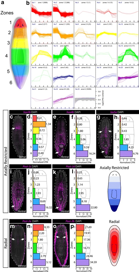
Fig. 2.

RNAseq and WISH reveal axially restricted HOX genes. a A cartoon schematic of 6 axial zones of tissue subjected to RNAseq is shown at the left. b Following transcript normalization across all zones, transcripts were binned according to having specific expression in particular zones. Twenty-two binning categories were distinguished, of which 16 contain specificity to more than one zone. For example, a specificity of 1.2 would signify that there was specificity to zones 1 and 2, with higher expression in zone 1 (whereas 2.1 has the same specificity but higher expression in zone 2). All transcripts and categories are listed in Additional file 3: Table S2. c–p FISH stains for individual HOX genes are shown in magenta and counterstained with DAPI in gray. Arrows denote where the strongest area of expression was seen. For each gene, the raw CPM values per zone are given in the histogram to the right of each stain. Two main categories were observed: axial restriction perpendicular to the A–P axis (c–l) and radial expression inside of the body edge at the D-V boundary (m–p)
