
Figure 2.
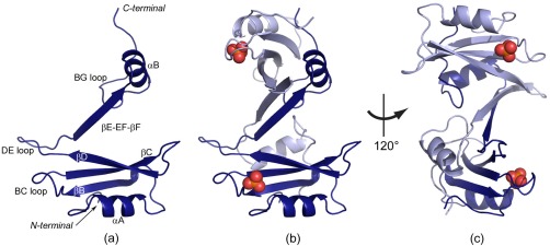
The Fyn SH2 dimer. (a) The monomer unit of the SH2‐dimer crystal structure with the main secondary structure elements labelled. The extended βE‐EF‐βF region and the αB‐helix adopt a different conformation than in the canonical SH2 fold. (b,c) The structure of the dimer in two orientations. The two monomer units are colored in light and dark blue, respectively. The cocrystallized phosphate is shown as spheres (phosphorus atoms are colored orange, oxygen atoms in red).