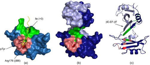
Figure 6.

Human Fyn SH2 free and in complex with the specific‐pY peptide. (a) The crystal structure of the Fyn SH2:specific‐pY complex. The main regions forming the pTyr binding pocket are colored in light red, while the specificity pocket is shown in green. The highly conserved Arg 176 (βB6) is labelled and illustrated in red. The specific‐pY peptide is colored in yellow and only the pTyr (0) and Ile (+3) residues are shown in sticks. (b) Surface and (c) cartoon representations of the Fyn SH2 dimer. The two monomer units are illustrated in light and dark blue, while the two binding pockets are shown for only one monomer unit using the same color codes as in (a).
