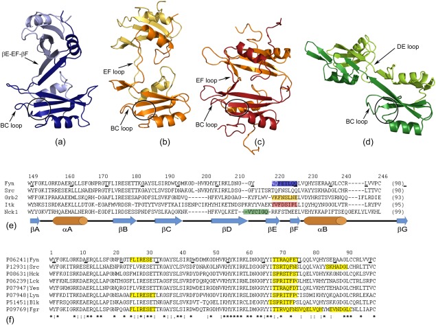
Figure 8.

Different SH2 dimers originating in (a) Fyn (PDB code: 4U17); (b) Grb2 (PDB code: 1FYR; Schiering et al., 2000); (c) Itk (PDB code: 3S9K; Joseph et al., 2012) and (d) Nck1 (PDB code: 2CI8; Frese et al., 2006). The monomer units for each SH2 domain are illustrated in dark and light color and are shown in the same structure‐orientation. The BC loop and the extended hinge region are labeled in each dimer, while the pTyr binding pocket is indicated by a black circle. (e) A multiple sequence alignment of SH2 domains from human Fyn, human Src, human Grb2, mouse Itk, and human Nck1. The disposition of the hinge regions is highlighted in the corresponding color as in (a–d). The residue numbers of human Fyn SH2 and the secondary structure elements are indicated. (f) A multiple sequence alignment of Src‐related SH2 domains highlighting the regions having the potential to form amyloid aggregates.
