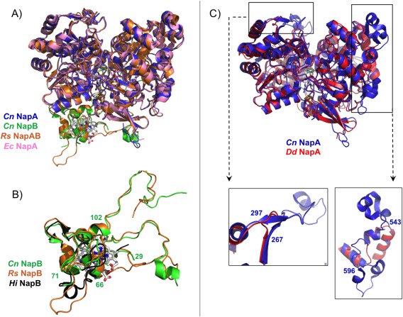
Figure 5.

Superpositioning of the crystal structures of the periplasmic nitrate reductases deposited in the PDB: (A) Comparison between heterodimeric proteins: NapAB from C. necator (NapA in blue and NapB in green, PDB ID: 3ML1), NapAB from R. sphaeroides (orange, PDB ID: 1OGY), and NapA from E. coli (pink, PDB ID: 2NYA). (B) Highlight of the comparison between Cn (green), Rs (orange), and Hi (black, PDB ID: 1JNI) small subunit NapB, with evidence of the similarities in the core structure of the three proteins (Cn NapB numbering). (C) Comparison between the catalytic subunits from Cn NapAB and Dd NapA, with a closeup of the two loops that differ considerably between heterodimeric and monomeric proteins (Cn NapAB numbering).
