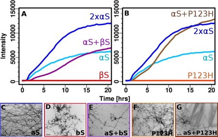
Figure 3.

Aggregation inhibiton of αS by βS and aggregation enhancement of αS by P123H‐βS (A & B). ThT fluorescence (37°C with shaking and teflon beads, PBS) of αS coincubated with (A) βS and (B) P123H‐βS. Negatively stained electron micrographs (scale 200 nm) (C‐G) of (C) αS fibrils, (D) βS amorphous aggregates, (E) coincubated αS with βS, (F) P123H‐βS amorphous aggregates, and (G) coincubated αS with P123H‐βS.
