
Figure 4.
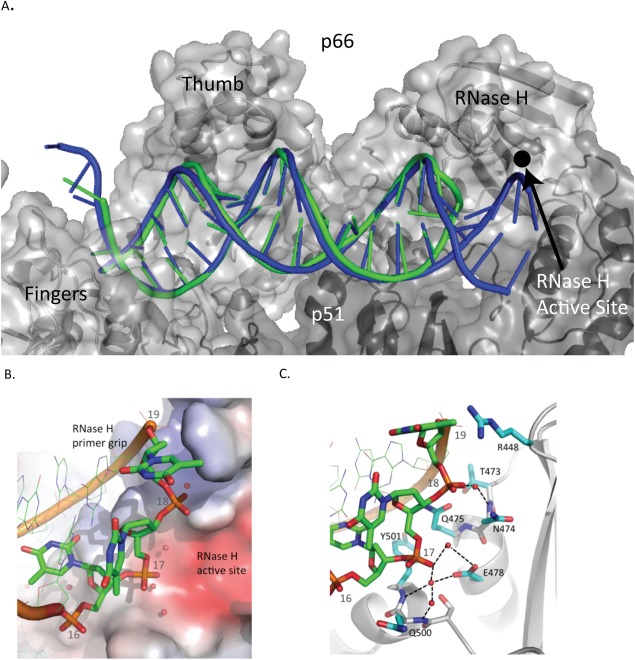
Comparison of the RT/aptamer structure (green) with the RT/cross‐linked‐dsDNA structure (3V6D) (blue). A. Nucleic acid binding cleft with the polymerase active site at the left and RNase H active site on the right. B. Aptamer hairpin (green) and cross‐linked‐dsDNA template strand (blue). Cross‐linked‐dsDNA template strand shown extending into RNase H active site. C. Aptamer binding pocket shown with selected side chains that are within 3.8 Å of nucleic acid atoms. A water molecule, shown as a red sphere, forms hydrogen bonds with Glu478 and the phosphate of T17.