
Figure 2.
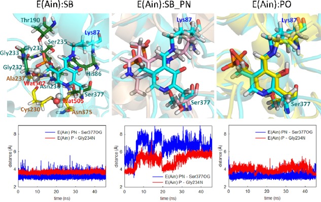
Protonation states of E(Ain). (left) The ligand (cyan) shows a proton at the SB nitrogen. The residues and water molecules forming interactions with E(Ain) are also shown. (middle) A proton was added at the pyridine nitrogen (PN). Motions of the ligand with two different protonation states, SB (cyan) and SB_PN (pink). (right) Comparison of a proton at the PO (yellow) and SB nitrogen (cyan). The distance between the PN and Ser377Oγ is shown in blue, and the distance between the phosphorous atom and Gly234N is shown in red. All snapshots are from 50‐ns MD simulation.