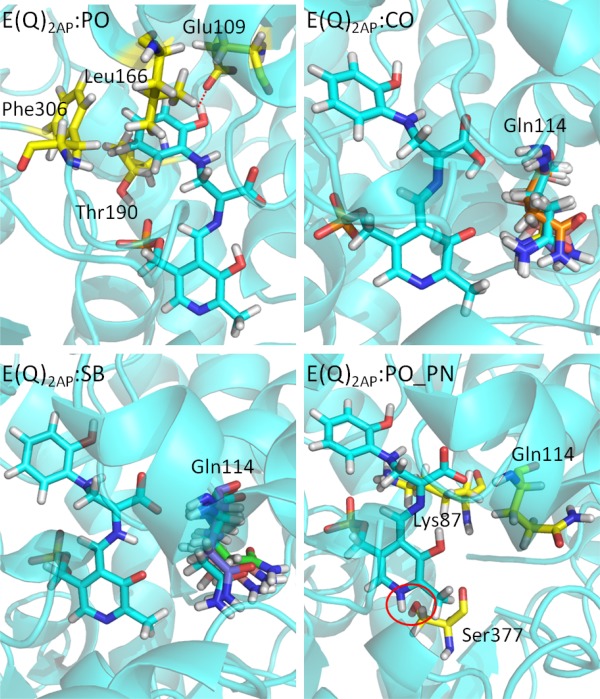
Figure 7.

Protonation states of E(Q)2AP. (PO) The residues interacting with the 2‐aminophenol ring of E(Q)2AP:PO are shown in a yellow bond representation. (CO) and (SB) The residue Gln114 is flexible during the 50 ns MD simulations. The side chain of Gln114 can move toward either the PO or solvent depending on protonation state. The conformations of Gln114 at 10, 20, 30, 40, and 50 ns are shown in different colors. (PO_PN) The key residues around E(Q)2AP:PO_PN are shown in yellow bond. The red circle indicates the missing H‐bond between the PN and Ser377. All snapshots are from 50‐ns MD simulation.
