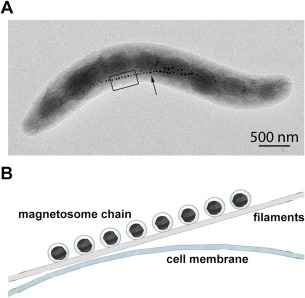
Figure 1.

Magnetotactic bacterium. (A) Transmission electron microscope (TEM) image of Magnetospirillum gryphiswaldense MSR‐1, contributed by Dr. René Uebe and Dr. Dirk Schüler. The black arrow points toward the magnetosome chain. (B) Magnified illustration of the black box in A: magnetosomes are made of magnetic particles surrounded by a lipid membrane—which invaginate from the cell membrane—and organized as a chain on filaments.
