Figure 3.

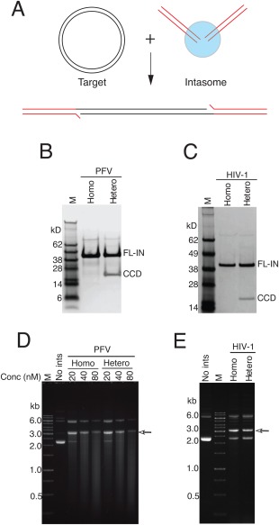
Activity of heterointasomes. A: Assembled intasomes were mixed with a circular target DNA in the presence of Mg2+. The integration product is linearized target DNA flanked by a pair of viral DNA ends. B: SDS‐PAGE of PFV heterointasomes. C: SDS‐PAGE of HIV heterointasomes. D: Activity of PFV heterointasomes. E: Activity of HIV heterointasomes. The final concentration of intasomes in the reaction mixture was 20 nM. The arrow indicates the migration position of the concerted integration product in agarose gel electrophoresis.
