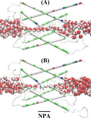
Figure 3.

Illustration of binding means inhibition. Single file waters in the apo state versus water line interrupted by PDO in the holo state of the PDO‐AQP2 complex. Protein is represented as cartoons colored by residue types; PDO as licorices colored by atom names; and waters inside the channel and near the channel entry/exit as large spheres colored by atom names. Color schemes: hydrophilic, green; hydrophobic, white; negatively charged, red; positively charge, blue; hydrogen, white; oxygen, red; carbon, cyan (All molecular graphics in the article were rendered with VMD.59).
