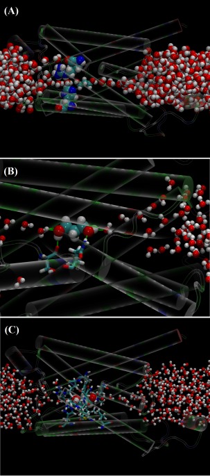
Figure 4.

PDO at the binding site near the NPA motifs. (A) Protein in cartoons colored by residue types, ar/R selectivity filter Arg 187 and His 172 in large spheres colored by atom names, PDO and water in or near the channel in medium spheres colored by atom names. This panel shows PDO residing near the NPA motifs which is on the right‐hand side of the ar/R and where the two half membrane helices face each other. (B) Zoom‐in of PDO with near‐by residues (Ser 182 and Asn 184) and waters forming four hydrogen bonds (green dashed bars). (C) PDO (large spheres colored by atom names) in favorable (attractive) van der Waals contact with surrounding residues (licorices colored by atom names). Color schemes identical to Figure 3.
