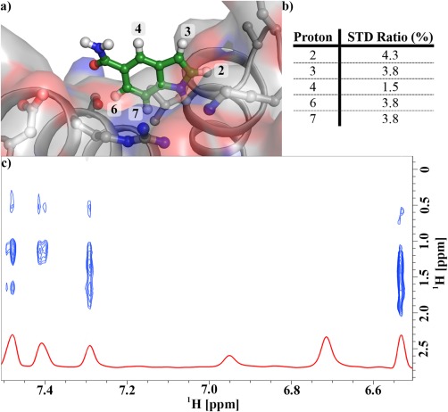
Figure 5.

Dock of Cp‐α1 into the 3S ΔF508 NBD1 structure is consistent with chemical shift perturbation and STD data. (a) The dock of Cp‐α1 into the α‐site composed of the apex of helices α4, α5, and α6. (b) STD ratios for the compound protons. STD signal strength is a function of distance from the protein; the protons closest to the protein have the highest STD ratios. (c) NOEs between Cp‐α1 protons and unassigned NBD1 methyl protons.
