Figure 4.

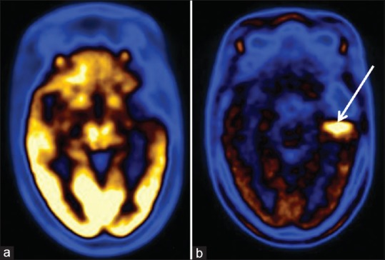
Grade I pilocytic glioma in left temporal lobe after surgery, radiotherapy, and chemotherapy. (a) Fluorodeoxyglucose scan was negative. (b) C-11 methionine scan shows residual/recurrent lesion in left temporal region

Grade I pilocytic glioma in left temporal lobe after surgery, radiotherapy, and chemotherapy. (a) Fluorodeoxyglucose scan was negative. (b) C-11 methionine scan shows residual/recurrent lesion in left temporal region