Figure 5.

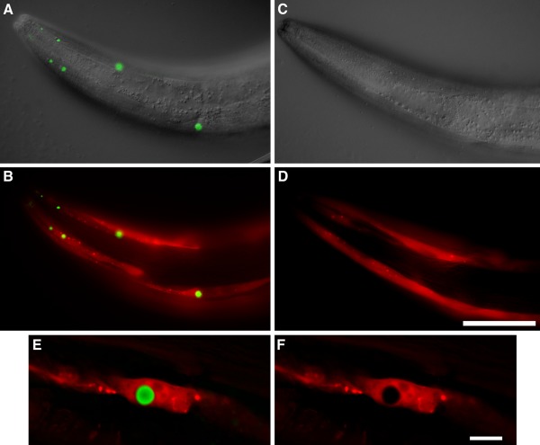
Poly Q inclusions detected with DCVJ. Transgenic strains expressing either Punc‐54::Q40 + Pmyo‐3::mCherry (CL1781) or Pmyo‐3::mCherry only (CL6617) were stained in parallel with DCVJ, and DCVJ fluorescence was imaged using identical exposure settings for the two strains. A: CL1781, overlay of DIC and DCVJ images. B: CL1781, DCVJ and mCherry fluorescence. C: CL6617, overlay of DIC and DCVJ images. D: CL6617 DCVJ and mCherry fluorescence. E: Higher magnification image of single DCVJ‐positive inclusion in strain CL1781, single optical section from digitally deconvolved image. F: Same as (E), mCherry channel only. Note: DCVJ only detects Q40 inclusions. Size bar = 50 μm, panels A−D; = 10 μm, panels E and F. DIC and epifluorescence images were acquired on a Zeiss Axiophot compound microscope equipped with a computer‐controlled Z‐drive and software from Intelligent Imaging Innovations. Photoshop software (Adobe) was used to fuse DIC and epifluorescence images.
