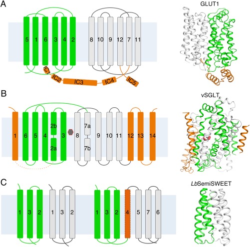
Figure 2.

The structural folds and overall structures of glucose transporters. (A) Structure of GLUTs. The 12 transmembrane helices were divided into an N domain and a C domain, which are colored green and white, respectively. The intracellular helices domains (ICH) are colored orange. Shown on the right is the structure of the human GLUT1 (PDB accession code 4PYP). (B) Structure of SGLTs. The transmembrane helices TM2‐11 constitute the “5 + 5” inverted repeats of the LeuT‐fold of vSGLT. The two repeats are colored green and white. The additional transmembrane helices are colored orange. The substrate (galactose) is indicated by the black hexagon. Shown on the right is the structure of the inward‐occluded vSGLT (PDB accession code 3DH4). (C) Structure of SemiSWEETs. The topology of SemiSWEETs and predicted topology of SWEET1 in human are shown on the left. All SemiSWEETs form a functional dimer. Each protomer contains three transmembrane helices, which are arranged in a 1–3–2 pattern and colored as white or green. Shown on the right is the structure of LbSemiSWEET (PDB accession code 4QNC). In all the figures where overall structures are shown, the cytoplasmic side is at bottom if not otherwise indicated.
