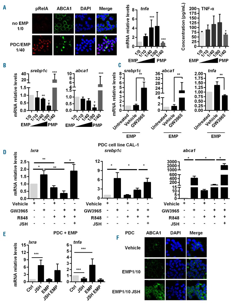
Figure 3.

NF-κB triggering by EMP stimulation or TLR7 activation prevents LXR activation in PDC. (A) Freshly isolated PDC were incubated with EMP at different PDC/EMP ratio (i.e. without, 1/0; one PDC for 10 EMP, 1/10; one PDC for 40 EMP, 1/40; one PDC for 80 EMP, 1/80). (Left) Nuclear translocation of phosphorylated NF-κBp65 (pRelA) subunit was determined in PDC cultured at a 1/40 ratio for 2.5 h by confocal microscopy. Nuclei were stained with DAPI. EMP stimulation of PDC decreases also membrane ABCA-1 expression. Results from a representative experiment out of 3. (Middle) tnfa mRNA levels from PDC cultured with EMP at the different ratio or with PMP (1/40 ratio) for 18 h, were quantified by qRT-PCR (n=5). (Right) TNF secretion from PDC cultured with EMP at the different ratio or with PMP (1/40 ratio) for 24 h were quantified by multiplex assays (n=5 for EMP and n=3 for PMP). (B) PDC were cultured with EMP at different PDC/EMP ratio (i.e. without, 1/0; one PDC for 10 EMP, 1/10; one PDC for 40 EMP, 1/40; one PDC for 80 EMP, 1/80) for 18 h. LXR target gene (srebp1c, abca1) mRNA transcripts were quantified by qRT-PCR (n=5). (C) PDC were treated with the LXR agonist GW3965 (1 μM) or vehicle (DMSO) for 24 h. Then, cells were washed and cultured with EMP at a 1/40 PDC/EMP ratio for 18 h (EMP). Untreated PDC were used as control. Expression of LXR target genes (lxra, abca1), and tnfa gene were quantified by qRT-PCR (n=5). (D) Cells from PDC cell line CAL-1 were treated with GW3965 (1 μM), R848 (1 μg/mL), JSH-23 (JSH, 25 μM), or a combination of these treatments for 18 h. LXR target gene (lxra, srebp1c, abca1) mRNA transcripts were quantified by qRT-PCR (n=5). (E) Freshly isolated PDC were treated for 18 h with the NF-κB inhibitor, JSH-23 (JSH), with or without EMP at a PDC/EMP ratio of 1:40. Expression of LXR target gene (lxra) and inflammatory cytokine [tnfa, il6 (not depicted)] gene mRNA was quantified by qRT-PCR (n=3). (F) PDC were treated with the NF-κB inhibitor JSH-23 (JSH) with or without EMP (at a PDC/EMP ratio of 1/10, EMP/1/10) for 2.5 h. Membrane ABCA1 expression was analyzed by confocal microscopy (one representative experiment out of 3 using PDC from 3 different donors). Data from n independent experiments were expressed as mean±S.E.M. *P<0.05, **P<0.001, ***P<0.005 (Mann-Whitney).
