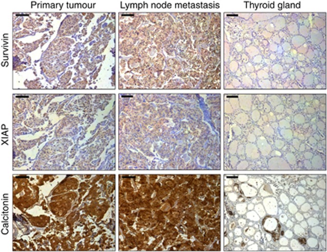
Figure 1.
Representative tissue samples with immunohistochemical staining for survivin, XIAP and calcitonin in MTC (left), respective lymph node metastases (middle) and normal thyroid gland (right). All shown samples were classified as strong expression for the respective targets in accordance with the IRS. The bar at the top left corner indicates 50 μm.

