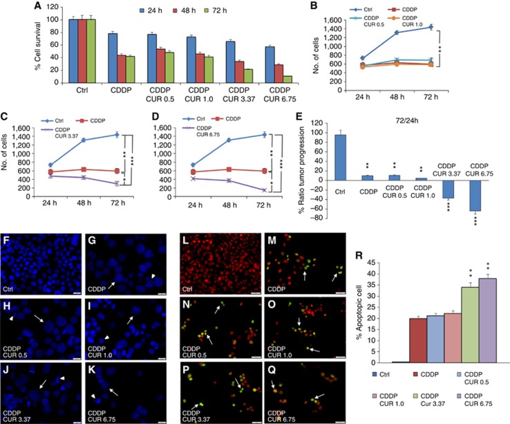
Figure 2.
Curcumin increases cisplatin effect in the combined administration. (A–D) Graphs (mean±s.e.m.) showing percentage of cell survival (A) and proliferation (B–D) at 24, 48 and 72 h of incubation with cisplatin and curcumin at different doses. (E) Histogram shows the tumour progression between 24 and 72 h, expressed as a percentage of the ratio of the number of cells counted at 72 and 24 h. *P<0.05, **P<0.001 and ***P<0.0001. (F–K) Representative images of DAPI-stained cells; arrows and arrowheads indicate swelling and nuclear condensation/cellular fragmentation. Scale bar: 20 μm. (L–Q) Terminal deoxynucleotidyl transferase dUTP nick-end labeling (TUNEL) assay showing marked apoptotic-positive cells after cisplatin treatment (M) and after an adjuvant curcumin treatment (N–Q). (R) Related apoptotic cell count (mean±s.e.m.). **P<0.001. Data are representative of three independent experiments; each count performed on 10 fields randomly selected for each experimental condition and each time point.

