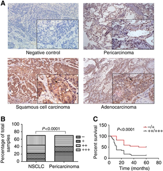
Figure 2.
Enhanced TIM-4 expression in NSCLC shows poor prognosis. Immunohistochemical (IHC) staining was performed in 70 cases of NSCLC tissues and pericarcinoma tissues. (A) Representative IHC staining images of TIM-4 in NSCLC tissues and pericarcinoma issues (× 200). The negative control indicated that rabbit IgG replaced TIM-4 antibody in the process of IHC staining. (B) Staining intensity of TIM-4 in NSCLC tissues was significantly higher than that of the pericarcinoma tissues (P<0.0001). (C) The survival rate of lung cancer patients with higher TIM-4 expression was significantly lower than that of the patients with lower TIM-4 expression (P<0.0001).

