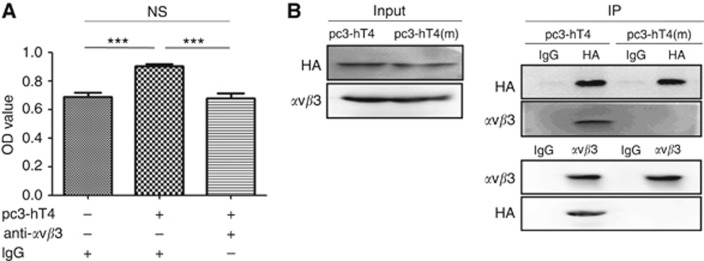
Figure 4.
T-cell immunoglobulin domain and mucin domain 4 (TIM-4) interacts with integrin αvβ3 through its RGD motif. The A549 cells were seeded in 96- or 6-well plates. After grown to 80–90% confluence, the cells were transfected with pc3, pc3-hT4 or pc3-hT4(m) plasmid DNA separately. After transfection, αvβ3 blocking or Co-IP was performed at indicated time points. (A) At 6 h after transfection, 25 μg ml−1 of anti-αvβ3 or mouse IgG was added into cells and incubated for 4 days. The cell growth was monitored by CCK-8 assay (***P<0.001). (B) At 48 h after transfection with pc3-hT4 or pc3-hT4(m) plasmid DNA, Co-IP assay was performed. These experiments were repeated at least three times.

