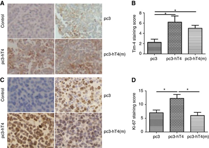
Figure 7.
T-cell immunoglobulin domain and mucin domain 4 (TIM-4) and Ki-67 expression in xenograft tumour tissues. The A549 cells were subcutaneously injected into the left axillae of the nude mice, and the tumours were injected with plasmid DNA of pc3, pc3-hT4 or pc3-hT4(m) every fourth day for a total of 4 injections. After 14 days, the mice were killed and the tumours tissues fixed in 10% buffered formalin were embedded in paraffin for IHC staining. The control represented the isotype control IgG instead of TIM-4 antibody or Ki-67 antibody in the process of IHC staining. (A) The TIM-4 and (C) Ki-67 protein expression in xenograft tumour tissues was examined by IHC staining. Photos of IHC staining are representative of at least 10 similar observations (× 200). The expression rate and staining intensity of (B) TIM-4 and (D) Ki-67 were reported separately according to the German semiquantitative scoring system (*P<0.05).

