Figure 6.
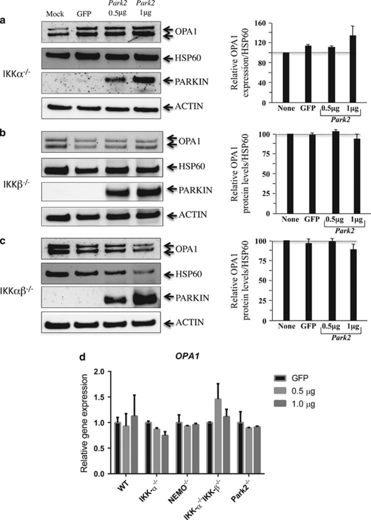
OPA1 expression in Parkin-transfected MEF cells. (a) IKKα−/−, (b) IKKβ−/−, and (c) IKKαβ−/−. MEFs were cotransfected with vectors encoding PARK2, GFP, or mock. The values indicate the relative levels of the long isoforms with respect to the baseline (value 1), in the absence (mock) or after 24 h of transfection with either GFP or PARK2 at the dose of 0.5 and 1 μg. HSP60 was used as a loading control to normalize protein content. Immunoblots were also probed with specific Parkin antibodies. Actin was used as a loading control. Four experiments were performed. (d) mRNA expression of OPA1 in Parkin–transfected MEF cells. RT-PCRs were performed in WT, IKKα−/−, NEMO−/−, IKKαβ−/−, and Parkin−/−. The values indicate the relative gene expression of OPA1 with respect to the GFP baseline (value set as 1). Two experiments were performed

