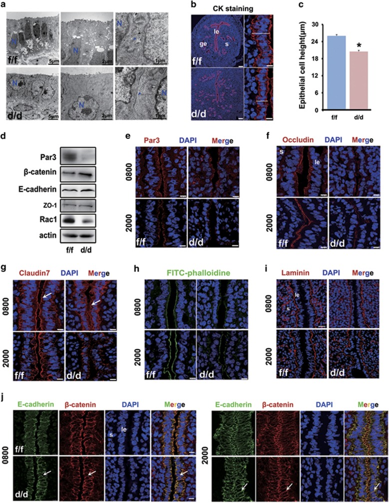
Figure 4.
Uterine epithelial transformation prior to implantation is disrupted in Rac1d/d mice. (a) Representative TEM pictures in Rac1f/f and Rac1d/d D4 uteri. N, cell nucleus. The blue arrowhead indicates cell-cell junction. (b, c) Measurement of luminal epithelial cell height by cytokeratin immunostaining in Rac1f/f and Rac1d/d D4 uteri. *P<0.05. The white line marks the length of the epithelium from basal to apical membrane. Scale bars for represents 100 and 10 μm, respectively. (d) Western blot detection of Par3, β-catenin, E-cadherin and ZO-1 in Rac1f/f and Rac1d/d D4 uteri. (e–h) Immunofluorescence localization of apical markers Par3, Occludin, Claudin 7 and F-actin in Rac1f/f and Rac1d/d uteri at D4 morning (0800 h) and evening (2000 h). The white arrowheads indicate apical staining in the luminal epithelium cells. Scale bars, 10 μm. (i) Immunostaining of basal membrane marker Laminin in Rac1f/f and Rac1d/d D4 uterus. Scale bars, 25 μm. (j) Immunofluorescence of adherens junction molecules E-cadherin and β-catenin in Rac1f/f and Rac1d/d D4 uteri. The white arrowheads indicate lateral membrane of the luminal epithelium cells. Scale bars, 10 μm. le, luminal epithelium; ge, glandular epithelium; s, stroma

