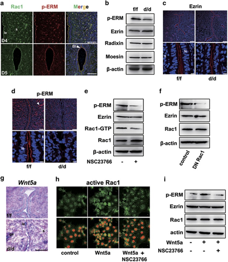
Figure 5.
ERM signaling is disrupted in Rac1d/d uteri. (a) Colocalization of Rac1 and p-ERM in Rac1f/f D4-5 uteri visualized by immunofluorescence staining. The white arrowhead indicates blastocyst. Scale bars, 50 μm. (b–d) Detection of p-ERM, Ezrin, Radixin and Moesin by western blot and immunofluorescence staining in Rac1f/f and Rac1d/d D4 uteri. The white arrowhead indicates blood vessel endothelium. Scale bars represent 100 and 10 μm, respectively. (e) Immunoblot analysis of p-ERM, Ezrin and Rac1 in Ishikawa uterine epithelial cells treated with 100 μM Rac1 inhibitor NSC23766 for 24 h. Actin serves as a loading control. (f) Western blot analysis of p-ERM, Ezrin and Rac1 in Ishikawa cells transfected with either the empty vector or dominant negative Rac1 (Rac1N17) constructs. Actin serves as a loading control. (g) In situ detection of Wnt5a in Rac1f/f and Rac1d/d mice. (h) Immunofluorescence detection of active Rac1 after Wnt5a and/or NSC23766 treatment. Scale bars, 50 μm. (i) Immunoblot analysis of Ishikawa cells treated with Wnt5a and/or NSC23766 for 24 h. Whole-cell lysates were blotted for p-ERM, Ezrin and Rac1. Actin serves as a loading control. le, luminal epithelium; s, stroma

