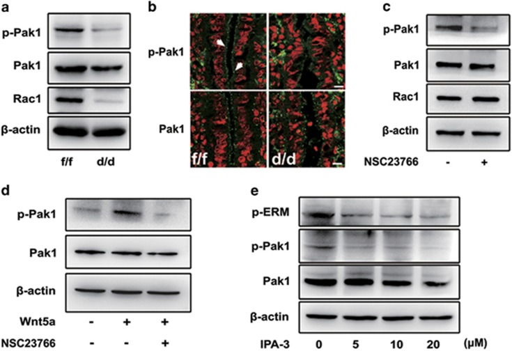
Figure 6.
Pak1 activation is inhibited in Rac1d/d luminal epithelium. (a, b) Western blot and immunostaining of p-Pak1 and Pak1 in the uterus of Rac1f/f and Rac1d/d mice. Actin serves as a loading control. White arrowheads indicate the positive signaling of p-Pak1. (c) Immunoblot analysis of p-Pak1, Pak1 and Rac1 in Ishikawa cells treated with Rac1 inhibitor NSC23766 for 24 h. Actin serves as a loading control. (d) Immunoblot analysis of Ishikawa cells treated with Wnt5a and/or NSC23766 for 24 h. Whole-cell lysates were blotted for p-Pak1 and Pak1. Actin serves as a loading control. (e) Immunoblot analysis of p-ERM, p-Pak1 and Pak1 in Ishikawa cells treated with various doses of Pak1 inhibitor IPA-3 for 24 h. Actin serves as a loading control

