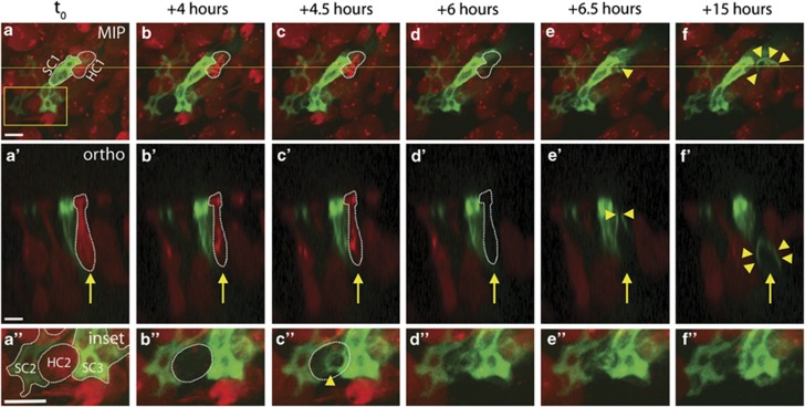
Figure 2.
SCs constrict the apical portion of dead or dying HCs before removing them from the sensory epithelium. (a–f) In a series of MIPs taken from a 48-h live-imaging experiment, a single HC (red, labeled HC1 in a) loses tdTomato signal at +6 h (dashed outline in d). The neighboring green SC (labeled SC1 in a) is observed constricting the apical portion of HC1 within 30 min of tdTomato loss (single arrowhead in e). SC1 then forms a phagosome surrounding the remains of HC1, eliminating the HC corpse from the sensory epithelium (multiple arrowheads in f). a'–f' Orthogonal views of the same imaging field as a–f, taken from the plane indicated by the yellow line in a. Arrow follows HC1, as it loses tdTomato (dashed outline, d') before SC constriction (arrowheads, e') and phagosome formation (multiple arrowheads, f'). a”–f” Magnified inset views taken from the outlined box in a. Two SCs (labeled SC1 and SC2 in a”) surround a single HC (labeled HC2 in a”), which loses tdTomato signal at +4 h (dashed outline, b”). SC2 and SC3 together constrict HC2 within 30 min (C”). Movies are Supplementary Figures 2 and 3. Overall gain was increased on inset images. Scale bars=5 μm

