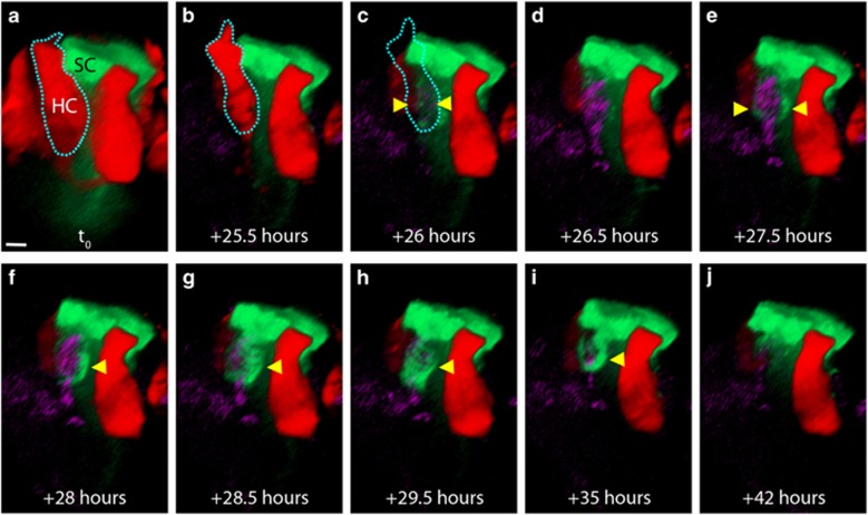
Figure 4.
The SC phagosome contains the nucleus of a dead HC. (a and b) 3D orthogonal view of a red HC and an adjacent green SC. (c) During live imaging, the HC loses tdTomato signal in the same frame in which TOTO-3 is weakly visible (arrowheads). (d) Thirty minutes later, the TOTO-3 is easily visible within the HC nucleus. (e): Within 1.5 h of tdTomato loss, the green SC has extended processes to surround the TOTO-3-positive nucleus (arrowheads). (f) A phagosome begins to surround the nucleus (arrowhead). (g and h) The SC phagosome has fully surrounded the nucleus. (i and j) The HC has been degraded and the phagosome appears to have receded. Movie is Supplementary Figure 8. Scale bar=3.5 μm

