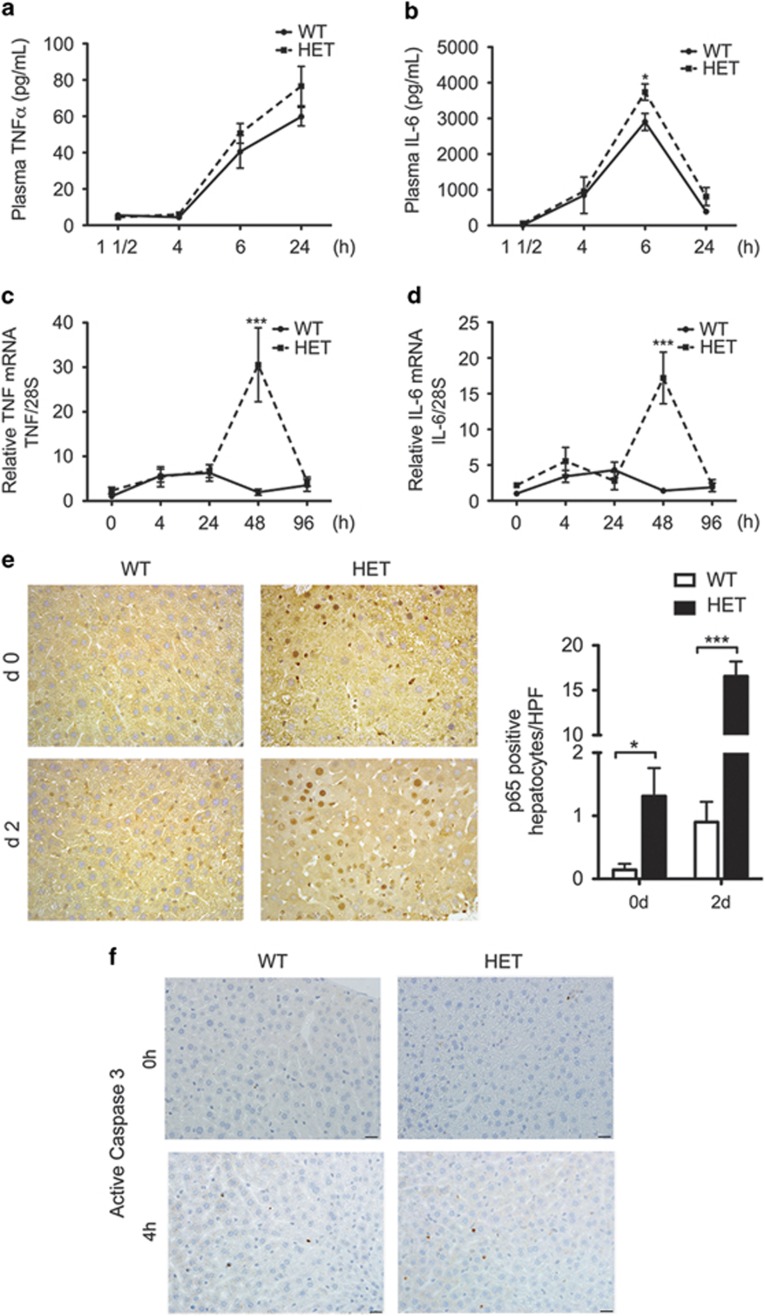
Figure 2.
A20 HET mice show higher intrahepatic levels of pro-inflammatory and priming cytokines, IL-6 and TNF, following PH. This correlates with increased activation of NF-κB in hepatocytes, but without aggravated apoptosis. Plasma samples were recovered from WT and A20 heterozygous (HET) mice before (0) and at several time points (1 0.5, 4, 24 h) after PH and evaluated by ELISA for plasma levels of (a) TNF and (b) IL-6. Livers from WT and A20 HET mice were recovered before (0) and 4, 24, 48 and 96 h after PH and evaluated for (c) TNF and (d) IL-6 intrahepatic mRNA levels by qPCR. Graphs represent mean±S.E.M. of mRNA levels relative to WT at time 0, respectively (n=3–8 mice per group). 28 S ribosomal (rRNA) was used as housekeeping gene. (e) Representative photomicrographs of p65 immunostaining (brown) before and 24 h after PH. Scale bars: 50 μm. Graph represents mean±S.E.M. of p65+ hepatocyte nuclei per high power field (HPF). Only p65+ nuclei that were considered hepatocytes based on size were counted (n=3 mice/group). *P<0.05, ***P<0.001. (f) Representative photomicrographs of active caspase 3 immunostaining (brown) before and 24 h after PH. Scale bars: 50 μm

