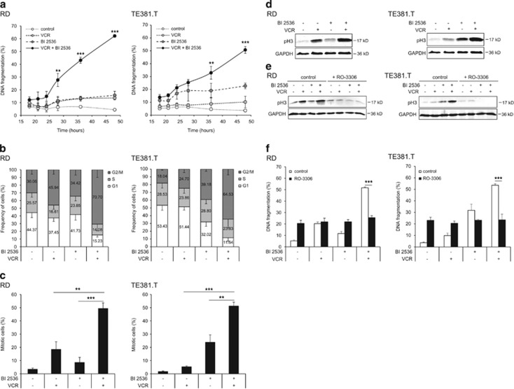
Figure 3.
BI 2536/VCR-induced mitotic arrest is required for apoptosis. RD cells were treated with 4 nM BI 2536 and/or 2 nM VCR, TE381.T cells with 7 nM BI 2536 and/or 1 nM VCR. (a) Apoptosis was determined at indicated time points by quantification of DNA fragmentation (n=3). (b) Frequency of cells per cell cycle phase was analyzed at 18 h in PI-stained nuclei using FlowJo software (TreeStar Inc.) (n=5). (c) Mitotic cells were quantified at 18 h by expression of mitotic marker pH3 using immunofluorescence (n=3). (d) pH3 expression was confirmed by western blotting at 18 h (n=3). GAPDH served as loading control and representative blots are shown. (e and f) RD and TE381.T cells were pretreated for one hour with 10 μM of CDK1 inhibitor RO-3306. pH3 expression at 24 h was confirmed by western blotting (n=3). GAPDH served as loading control and representative blots are shown (e). Apoptosis was determined at 48 h by quantification of DNA fragmentation (n=4). Results are expressed as mean±S.D. (error bars) (f). Student's t-test was used to calculate two-sided P-values. **P<0.01; ***P<0.001

