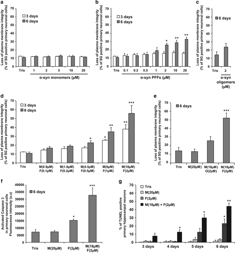
Figure 3.
Mixture of α-syn monomers and fibrils exacerbates cell death in hippocampal primary neurons. (a–f) Hippocampal primary neurons were plated in 96-well plate and treated with Tris buffer or α-syn species for the indicated times. (a–e) Cells were stained with Sytox Green (SG), a membrane-impermeant dye that will enter only in cells with damaged plasma membranes. Cell death level is expressed as the percentage of cells with compromised cell membrane (SG-positive cells) to the total cell number analyzed by Tecan infinite M200 Pro plate reader (487 nm/519 nm). (f) Caspase 3 activity was quantified by a caspase 3 activity assay using Tecan infinite M200 Pro plate reader (487 nm/519 nm). (g) Hippocampal primary neurons were plated on coverslips and treated up to 6 days with Tris buffer (negative control), α-syn monomers, sonicated α-syn PFFs or α-syn mixture. Cells were fixed at the indicated time with PFA 4% and subsequently stained for the apoptotic cells (TUNEL). Nucleus was counterstained using SG and the neuronal population was specifically stained using NeuN. The percentage of apoptotic neurons was quantified as follows: (TUNEL-positive and NeuN-positive cells)/total NeuN-positive cells. Each experiment included counting of three fields with an average of 400 cells/field per condition. (a–g) Data shown represent the means of three independent experiments performed in triplicate for each condition (bars are means±S.D.). One-way ANOVA test followed by Tukey–Kramer post-hoc test were performed (Tris versus α-syn-treated conditions), no statistical significance was observed in (a–c); *P<0.01, **P<0.001, ***P<0.0001 in (d, f and g)

