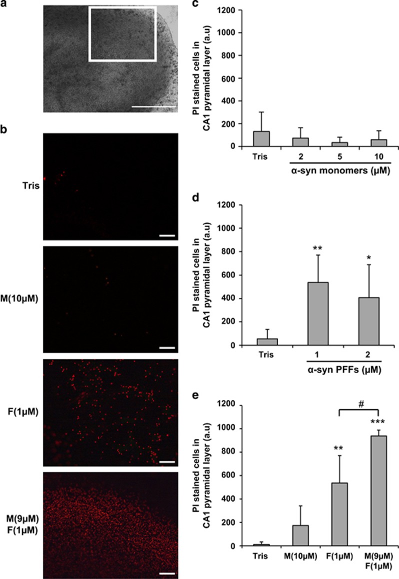
Figure 4.
Mixture of α-syn monomers and fibrils exacerbates cell death in ex vivo hippocampal slice culture. Hippocampal slices were treated at day in vitro (DIV) 13–14 with extracellular monomeric α-syn, sonicated α-syn PFFs or α-syn mixture. After 24 h, cell death was measured by quantification of PI-positive cells in the CA1 region. (a) Bright-field image of a hippocampal organotypic slice culture observed with a 5 × objective. Insert indicates CA1 region of analysis shown in (b). Scale bars= 500 μm. (b) Epifluoresence images of CA1 area acquired under a 10 × objective showing PI staining in different conditions of incubation: Tris, α-syn monomers (M; 10 μM), α-syn PFFs (F; 1 μM) and mixture of α-syn monomers and PFFS (9 μM M+1 μM F). Scale bars= 100 μm. (c) Quantification of PI-positive cells in CA1 pyramidal layer after incubation of Tris (n=52 cultures), α-syn monomers at 2 μM (n=39), 5 μM (n=7) or 10 μM (n=11). (d) Quantification of PI-positive cells in CA1 pyramidal layer after incubation of Tris (n=29), PFFs at 1 μM (n=8) or 2 μM (n=24). (e) Quantification of PI-positive cells in CA1 pyramidal layer after incubation of Tris (n=4), α-syn monomers 10 μM (n=4), PFFs 1 μM (n=8) and mixture of α-syn monomers (9 μM) and preformed fibrils (1 μM) (n=8). (c–e) Bars are means±S.D. *P<0.05, **P<0.001, ***P<0.0001 (Tris versus α-syn-treated conditions), #P<0.01 (α-syn PFFs versus α-syn mixture), one-way ANOVA followed by Tukey–Kramer post-hoc test

