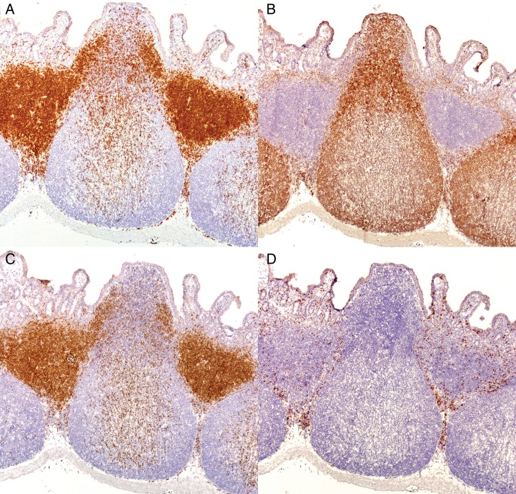
Figure 2.
Lymphocyte distribution in the ileocecal plaque of the rabbit (original magnification 100x). Immunohistochemistry of the ileocecal plaque with antibodies against (A) CD3, (B) CD79a, (C) CD4, and (D) CD8. The structure of the ileocecal plaque is similar to that of a Peyer's patch. (A) Anti-CD3 stains T cell–rich parafollicular regions as well as scattered cells throughout B cell–predominant follicles, the submucosa, and mucosa. (B) Inductive sites are flask shaped, consisting of CD79a positive follicles under a narrower CD79a positive subepithelial dome region. There is a predominance of (C) CD4 cells to (D) CD8 cells in all GALT effector sites.

