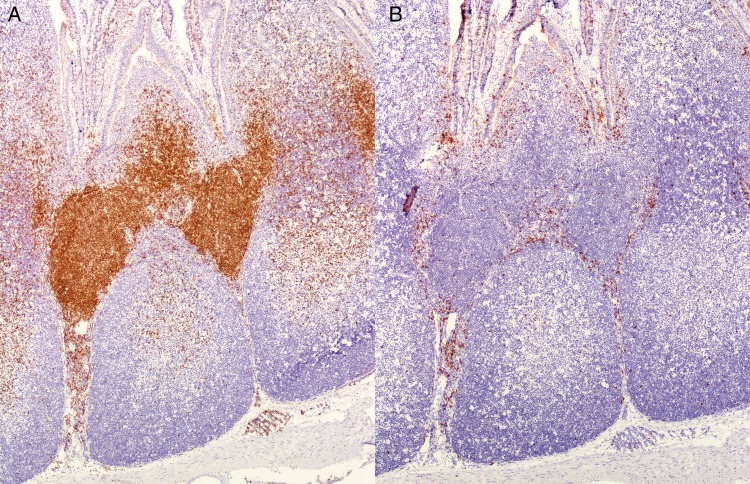
Figure 3.
CD4 and CD8 T cell distribution in cecal tonsil of the rabbit (original magnification 100x). Immunohistochemistry of the cecal tonsil with antibodies against (A) CD4 and (B) CD8 T cells. (A) Within T cell parafollicular zones, the majority of the cells are CD4 positive. T cell zones contain smaller numbers of (B) CD8-positive cells compared with (A) CD4-positive cells.

