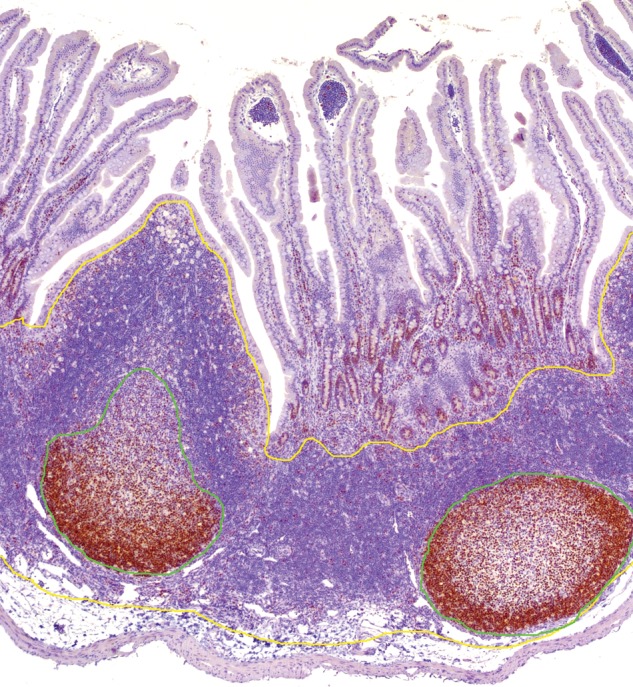
Figure 4.

Quantitative area analysis for Ki-67 in the Peyer's patch of the rabbit (original magnification 100x). Immunohistochemistry of the Peyer's patch stained with antibody against Ki-67. Positive staining identifies the germinal center. Formalin-fixed, paraffin-embedded tissue sections were stained with antibody and scanned (Aperio ScanScope XT; LeicaBiosystems). The cross-sectional area of the Peyer's patch, ileocecal plaque, cecal tonsil, and appendix were measured using the pen tool in Aperio ImageScope Viewer. The follicular region and germinal center (proliferation zone, green line) areas were outlined and calculated as a percentage of the total cross-sectional area (yellow line), as described in Dasso and colleagues 2000). The germinal center was defined as the area staining with Ki-67 (Dasso et al. 2000).
