Figure 1.
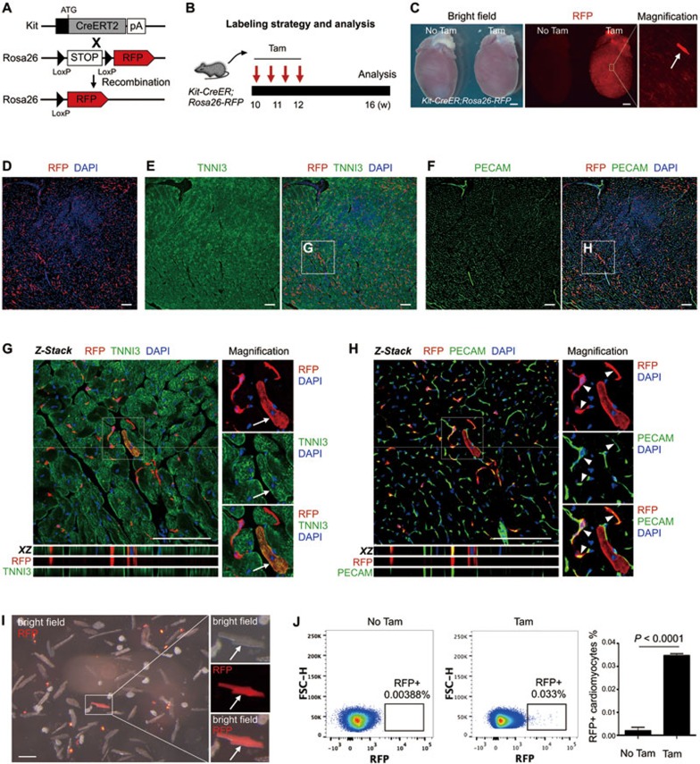
The fate of Kit+ cells in heart homeostasis. (A) Kit-CreER mice were crossed with Rosa26-RFP mice for lineage tracing. (B) A schematic of tamoxifen induction and analysis. (C) Whole mount bright field and fluorescence view of Kit-CreER;Rosa26-RFP hearts with or without tamoxifen (Tam or No Tam). Arrow indicates RFP+ cardiomyocyte. (D-F) Immunostaining for RFP, TNNI3 and PECAM on Kit-CreER;Rosa26-RFP heart sections. (G, H) Z-stack confocal images including XZ scanned sections showing RFP+TNNI3+ cells (arrows, G) and RFP+PECAM+ cells (arrowheads, H). (I) Image of cells dissociated from hearts of Kit-CreER;Rosa26-RFP mice. Arrow indicates a rare RFP+ cardiomyocyte. (J) Quantification of RFP+ cardiomyocytes by flow cytometry. Events in images are gated on live and lineage-negative cardiomyocytes. n = 4. Scale bars represent 1 mm in C, 100 μm in D-I.

