Figure 4.
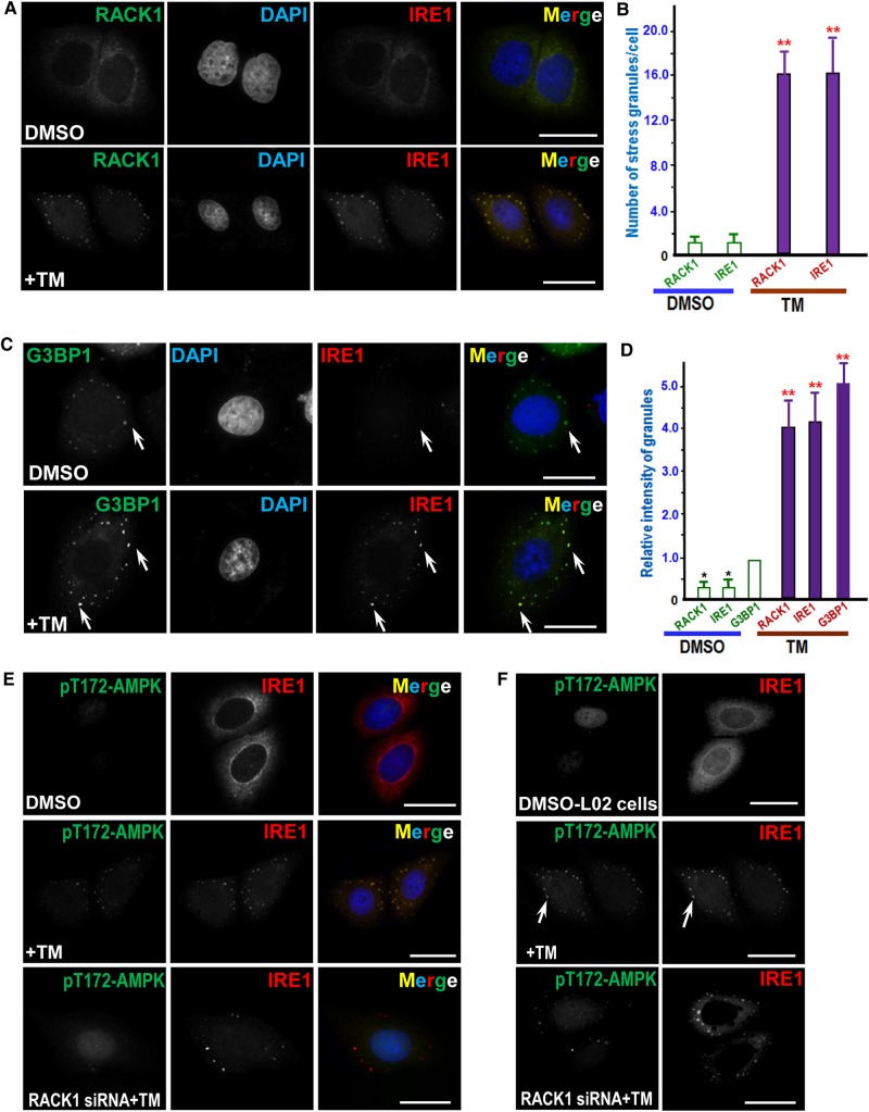
ER stress induced by TM treatment results in a RACK1-dependent co-localization of AMPK with IRE1. (A) Immunofluorescence of HepG2 cells probed for IRE1 and RACK1. TM-treated and control cells were fixed and permeabilized as described in Materials and methods, followed by probing with mouse IRE1 antibody and rabbit RACK1 antibody. IRE1 is visualized with a rhodamine-conjugated secondary antibody, while RACK1 is marked by a secondary antibody conjugated to FITC. Scale bar, 10 µm. (B) Quantitative analyses of stress-induced granules positive for IRE1 and RACK1 as a function of TM treatment. TM-treated and control cells were fixed, stained, and imaged as described in A. A total of 50 cells from three preparations were quantified. Error bars represent SEM. **P < 0.001 compared with DMSO-treated cells. (C) Immunofluorescence of HepG2 cells probed for IRE1 and G3BP1. TM-treated and control cells were fixed and permeabilized followed by probing with mouse IRE1 antibody and rabbit G3BP1 antibody. IRE1 is visualized with a rhodamine-conjugated secondary antibody, while G3BP1 is marked by a secondary antibody conjugated to FITC. Note that punctuate staining appears in TM-treated cells in which IRE1 and G3BP1 signals are super-imposed (yellow). Scale bar, 10 µm. (D) Quantitative analyses of stress granule intensity changes in response to TM treatment. TM-treated and control cells were fixed, stained, and imaged as described in C. A total of 50 cells from three preparations were quantified. Error bars represent SEM. **P < 0.001 compared with DMSO-treated cells. (E) Immunofluorescence of HepG2 cells probed for IRE1 and pT172-AMPK. IRE1 is marked by a rhodamine-conjugated secondary antibody, while pT172-AMPK is labeled by a secondary antibody conjugated to FITC. Aliquots of HepG2 cells were treated with RACK1 siRNA followed by TM treatment (bottom panel). Scale bar, 10 μm. (F) Immunofluorescence of L02 cells probed for IRE1 and pT172-AMPK. IRE1 is marked by a rhodamine-conjugated secondary antibody while pT172-AMPK is labeled by a secondary antibody conjugated to FITC. Aliquots of L02 cells were treated with RACK1 siRNA followed by TM treatment (bottom panel). Note that punctuate staining appears in TM-treated cells in which IRE1 and pT172-AMPK signals are super-imposed. Scale bar, 10 μm.

