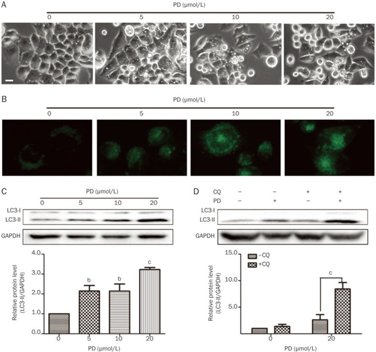
Figure 4.
PD triggers autophagy in BEL-7402 cells. BEL-7402 cells were treated with different concentrations of PD for 24 h. (A) The cell morphology was directly observed under an Axiovert 200 inverted microscope. Bar: 20 μm. (B) The autophagic vacuoles were labeled with MDC by incubating cells with 0.05 mmol/L MDC. (C) The protein levels of LC3 were determined by Western blot analysis. (D) The BEL-7402 cells were pretreated with 5 μmol/L CQ for 1 h followed by 20 μmol/L PD treatment for an additional 24 h, and the protein levels of LC3 were determined by Western blot analysis. cP<0.01 vs -CQ.

