Figure 4.
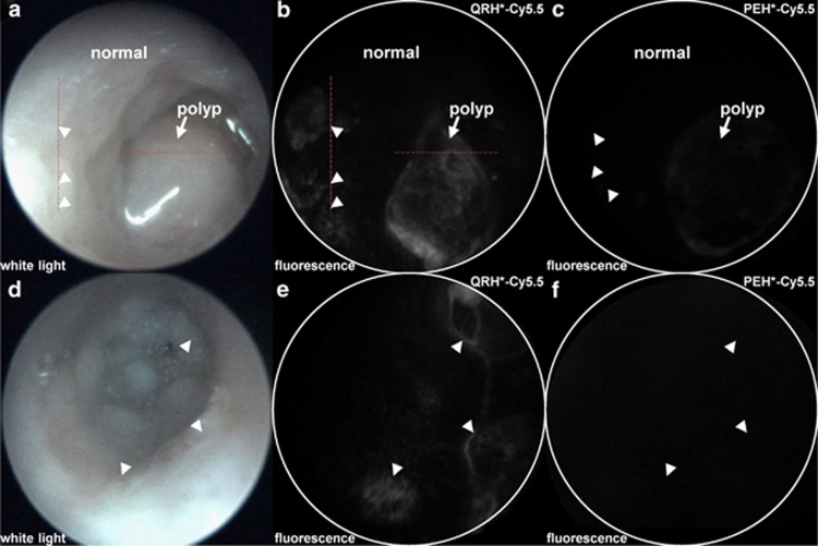
In vivo imaging of colon in CPC;Apc mouse. (a) White-light image of colon in CPC;Apc mouse shows the presence of polyp (arrow). Pathology was evaluated along (dashed) red lines. (b) NIR fluorescence image after topical administration of QRH*-Cy5.5 shows increased intensity from polyp (arrow) and several flat lesions (arrowheads) with heterogeneous pattern. (c) Image with PEH*-Cy5.5 shows minimal signal. (d) White-light image shows no grossly visible lesions (polyps). (e) NIR fluorescence image with QRH*-Cy5.5 shows the presence of flat lesions (arrowheads). (f) Image with PEH*-Cy5.5 shows minimal signal. NIR, near-infrared.

